
Weekly reads 22/6/26
How spatial context, summary statistics, and scalable frameworks are transforming biology

This week’s reads entail only 5 papers due to some well-deserved days away from my laptop, but the works are worth a read as they push the boundaries of spatial, computational, and functional genomics. SPACE-seq combines CRISPR lineage barcoding with spatial transcriptomics to identify intraclonal tumor heterogeneity, tumor-stroma interactions, and embryonic signatures in liver and brain. On the methods side, ReconEval evaluates how well single-cell latent spaces reconstruct gene expression and finds that simpler models like autoencoders often do better than their complicated counterparts. Rhaister, on the other hand, shows that one can predict drug and perturbation effects with excellent accuracy using only summary statistics, while SCALLOPS offers a scalable, cloud-based pipeline to handle terabyte-sized image datasets from optical pooled screens. Moreover, TIDEST proposes a statistical test for differential expression in spatial transcriptomics that accounts for prediction uncertainties and spatial confounding.
Preprints/articles that I managed to read this week
SPACE-seq integrates spatial transcriptomics and lineage tracing in native tissues
Jia et al., Cell Stem Cell (2026). 10.1016/j.stem.2026.05.017
The paper in one sentence
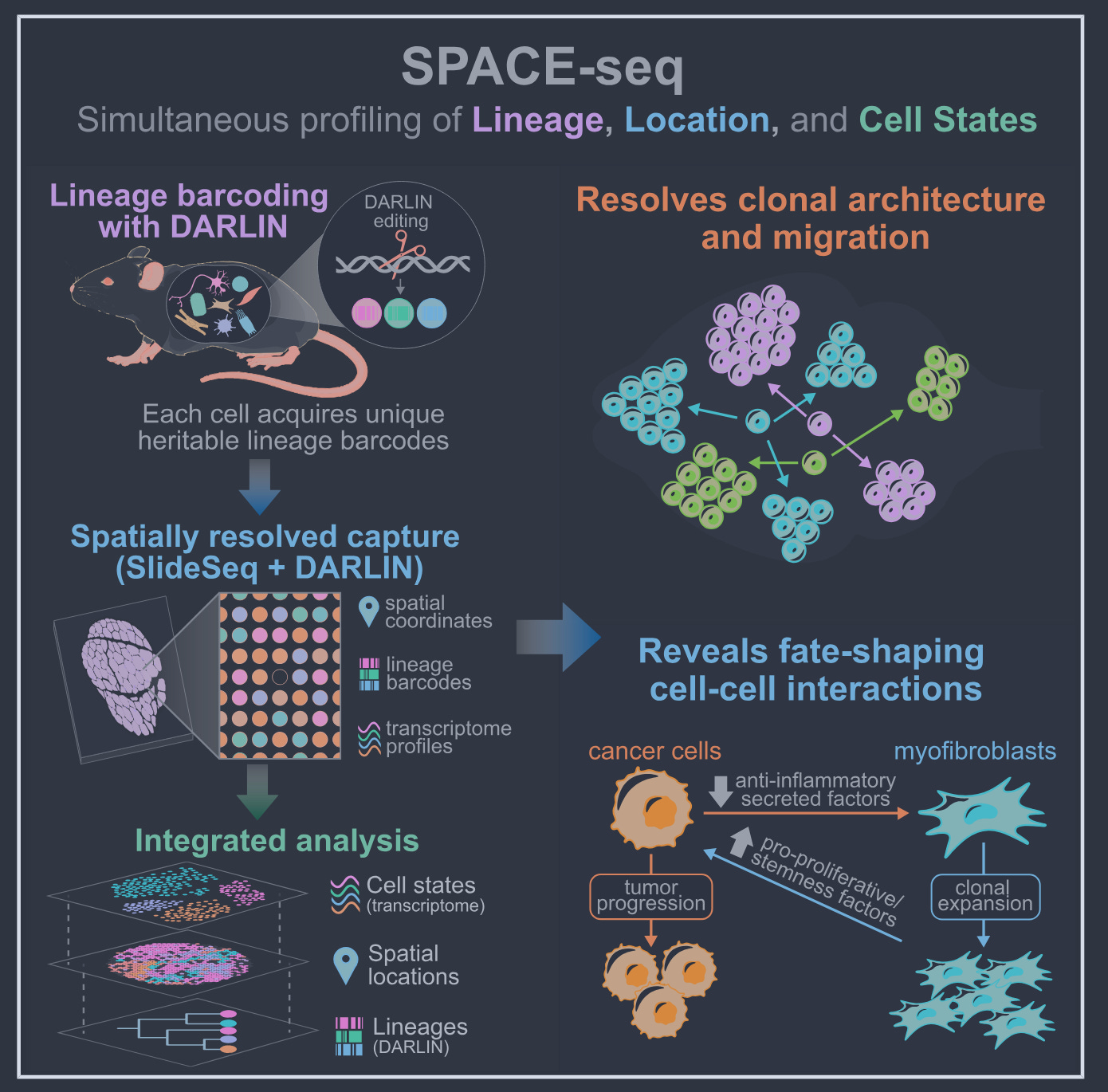
This study introduces SPACE-seq, a technology that integrates CRISPR-based lineage barcoding with spatial transcriptomics to jointly map cellular ancestry, identity, and location in intact tissues at near-cellular resolution.
Summary
The authors developed SPACE-seq to address a long-standing limitation in biology: the inability to simultaneously track cellular lineage, transcriptional state, and spatial position within native tissues. By combining the DARLIN barcoding system (a CRISPR-based lineage recorder) with Slide-seqV2 spatial transcriptomics, they achieved multimodal profiling that preserves spatial context. The platform’s capabilities are demonstrated through applications in liver cancer (iCCA), brain development, and liver organogenesis, revealing new insights into clonal architecture, cell migration, and fate-shaping cellular interactions that were previously inaccessible. The study also validates the functional relevance of these findings, such as the role of SELENOP in suppressing fibrosis, using mouse models and human datasets.
Personal highlights
Multimodal spatial-lineage mapping: SPACE-seq integrates DARLIN CRISPR barcoding with Slide-seqV2, enabling joint profiling of lineage (via heritable barcodes), whole-transcriptome, and spatial coordinates at 10 μm resolution.
Intraclonal tumor heterogeneity: In an NRAS/p53-driven iCCA model, clonally related tumor cells diversify into distinct transcriptional states (e.g., differentiated vs. progenitor-like), supporting plasticity rather than distinct cells of origin as a driver of tumor heterogeneity.
Tumor-stroma crosstalk: Identifies bidirectional interactions between tumor cells and hepatic stellate cells (HSCs), with tumor-derived SELENOP acting as a fibrosis-suppressive factor. This axis is functionally validated in mouse models and correlated with human cholangiocarcinoma and NAFLD datasets.
Developmental window for liver patterning: Reveals a restricted developmental window (before E12.0 in mice) during which hepatoblasts disperse broadly to establish spatial domains that prefigure liver lobar architecture, challenging previous assumptions about liver lobe formation.
3D lineage reconstruction: Demonstrates integration of consecutive tissue sections to reconstruct 3D lineage maps, uncovering symmetric clonal distributions in hippocampal subregions and providing a framework for studying complex tissue organization.
Why should we care?
This work provides a significant technical advance by enabling the simultaneous measurement of three critical dimensions of cell biology: lineage, state, and space. The findings challenge the "distinct cell-of-origin" model for iCCA heterogeneity by demonstrating intraclonal diversification, suggesting that tumor plasticity may be a more significant driver of phenotypic diversity than previously appreciated. The discovery of the SELENOP-HSC axis as a regulator of fibrosis offers a concrete example of how spatial lineage information can uncover actionable biology, though its therapeutic potential remains to be rigorously tested in clinical settings.
Benchmarking gene expression reconstruction from single-cell latent representations
Fu et al. bioRxiv (2026). 10.64898/2026.06.15.731445
The paper in one sentence
This work introduces ReconEval, a benchmark that systematically evaluates how faithfully latent representations in single-cell genomics can reconstruct gene expression, revealing that simpler models often outperform complex alternatives for this critical but overlooked task.
Summary
Single-cell transcriptomics relies heavily on low-dimensional latent representations (e.g., PCA, autoencoders, VAEs, or foundation model embeddings) to denoise data, integrate datasets, and model cellular responses to perturbations. However, the ability to reconstruct biologically meaningful gene expression from these latent spaces—a prerequisite for interpreting predicted cell states—has lacked systematic evaluation. The authors address this gap with ReconEval, a benchmark assessing reconstruction quality across >100 million cells from three diverse datasets: Tahoe-100M (drug perturbations), PBMC-10M (cytokine perturbations), and LuCA (observational lung cancer atlas).
The study evaluates three reconstruction paradigms:
End-to-end reconstruction (PCA, autoencoders (AEs), VAEs) with joint encoder-decoder training.
Foundation-model reconstruction, where pretrained embeddings (from STATE, scConcept, scGPT, SCimilarity) are paired with separately trained decoders (MLP, KNN, Transformer).
Latent shift reconstruction, where perturbation prediction models (STATE, CellFlow) operate on latent spaces before decoding.
Metrics span statistical fidelity (MSE, R², energy distance), biological signal preservation (differential expression, pathway activity, coexpression, cell cycle), and perturbation-specific effects (KNN purity). Key findings include: AEs outperform VAEs and PCA for stand-alone reconstruction, especially at low dimensionality; foundation model embeddings retain decodable gene-level signal but vary in performance by pretraining objective and decoder architecture; and optimal latent spaces for perturbation prediction depend on the downstream model (e.g., high-dimensional PCA for STATE, low-dimensional AEs for CellFlow).
Personal highlights
Autoencoders as the default for reconstruction: AEs consistently outperform VAEs and PCA across datasets and out-of-distribution regimes, even at extremely low latent dimensions (d=10), challenging the assumption that variational regularization improves generalization for reconstruction tasks.
Foundation models retain but don’t dominate: Pretrained embeddings (e.g., STATE’s SE) contain recoverable gene-level signal, but only SCimilarity outperforms PCA at matched dimensionality, and decoder choice (MLP > KNN > Transformer) is as important as the embedding itself.
Latent dimensionality is model-dependent: For one-step generative models (STATE), high-dimensional representations (PCA-2048) excel, while flow-based models (CellFlow) prefer low-dimensional AE embeddings (d=10), highlighting that optimal latent space depends on the downstream modeling paradigm.
Reconstruction ≠ representation quality: Strong performance in stand-alone reconstruction does not translate to latent shift reconstruction, underscoring that biological interpretability must be explicitly validated for each use case.
Practical guidance for representation selection: The work provides actionable recommendations: use AEs for end-to-end reconstruction, MLPs for foundation-model decoders, and high-dimensional PCA or low-dimensional AEs for perturbation prediction, depending on the model class.
Why should we care?
The authors address a current blind spot in single-cell genomics with their work: the assumption that latent spaces used for downstream tasks (e.g., perturbation prediction, virtual cells) can be meaningfully decoded back to gene expression. The takeaways are both practical and conceptual:
Simpler is often better: Despite the hype around foundation models and VAEs, basic autoencoders and even PCA frequently outperform them for reconstruction tasks, especially when dimensionality is constrained. This suggests that complexity does not guarantee biological interpretability, and that simpler, transparent methods may be preferable for tasks requiring gene-level insights.
One size does not fit all: The optimal latent representation depends on the downstream application. For example, high-dimensional PCA works well for one-step perturbation models (STATE), while low-dimensional AEs are ideal for flow-based models (CellFlow).
Foundation models are not a silver bullet: While pretrained embeddings (e.g., from STATE or scGPT) retain gene-level information, their performance is highly dependent on decoder architecture and does not uniformly surpass traditional methods. This highlights a gap between their success in tasks like cell-type annotation and their utility for gene-level reconstruction.
Biological interpretability must be explicit: The study reveals that reconstruction quality is not inherently tied to representation quality. A latent space may excel at clustering or integration but fail to preserve the gene-level signals needed for tasks like differential expression analysis or pathway scoring. .
Back to basics: Observed statistics are sufficient to predict drug responses
Svensson et al. bioRxiv (2026). 10.64898/2026.06.09.731197
The paper in one sentence
Rhaister is a fast, interpretable model that predicts drug and perturbation responses across new biological contexts using only summary statistics from large-scale screens, often matching or exceeding the performance of more complex virtual-cell models.
Summary
This work introduces Rhaister, a modeling framework that predicts how cells, tissues, or patients will respond to drugs, cytokines, or genetic perturbations by operating directly on aggregated summary statistics (e.g., log2 fold changes, p-values, expression deltas) from large perturbation screens like Tahoe-100M or the newly created Emerald Bay dataset. Unlike virtual-cell models that simulate single-cell data, Rhaister learns how response patterns vary across reference contexts and expresses unseen perturbations as linear combinations of observed ones. On transcriptional and phenotypic endpoints, Rhaister achieves state-of-the-art performance, often saturating evaluation metrics, while training in seconds and running predictions in milliseconds. The authors also present Rhaister-O, the first zero-shot model to predict drug responses in new contexts using only baseline gene expression, outperforming prior state-of-the-art models for this task.
Personal highlights
Summary-statistic modeling: Rhaister predicts perturbation responses directly from aggregated readouts (e.g., log2 fold change, p-values), bypassing the need for single-cell simulation and enabling rapid, interpretable predictions.
Performance at scale: On Tahoe-100M and Emerald Bay, Rhaister matches or exceeds virtual-cell models (e.g., STATE) across six evaluation metrics, often reaching half-sample reference performance, indicating saturated predictive accuracy.
Emerald Bay dataset: A new, publicly available perturbation atlas of cancer therapeutics applied to pooled Mosaic tumors, with paired drug sensitivity and transcriptomic readouts from 1.8M single cells, enabling context-specific drug phenotype prediction.
Zero-shot prediction with Rhaister-O: The first model to meaningfully predict perturbation responses in unseen contexts using only baseline expression data, outperforming complex baselines and prior state-of-the-art models.
Scalability and efficiency: Predictive performance improves with larger perturbation panels and more reference contexts, but even small panels (e.g., a single drug at three doses) can achieve near-optimal results with as few as 10 reference contexts.
Why should we care?
This work challenges the assumption that predicting biological responses requires simulating individual cells. By focusing on summary statistics, Rhaister demonstrates that fast, accurate, and interpretable predictions are possible without the computational overhead of virtual-cell models. Another takeaway is that large, diverse datasets (like Tahoe-100M and Emerald Bay) can be leveraged to predict drug responses in hard-to-study contexts (e.g., rare cell types, patient tissues) with minimal experimental input. This shifts the bottleneck from model complexity to data generation, making it feasible to integrate predictive modeling into high-throughput reasoning systems. Critically, while Rhaister excels at predicting pre-defined, static readouts, it may not capture emergent single-cell behaviors (e.g., novel cell types or synergistic gene interactions) that virtual-cell models aim to simulate. Its strength lies in practical, scalable prediction for downstream analysis.
SCALLOPS: a scalable, integrated computational framework for Optical Pooled Screens
Gould et al. bioRxiv (2026). 10.64898/2026.05.22.727250
The paper in one sentence
SCALLOPS is a unified, modular, and cloud-native computational framework that overcomes the main computational bottlenecks in processing terabyte-scale, multimodal image data from Optical Pooled Screens (OPS), enabling accurate genotype-phenotype linkage and scalable feature extraction.
Summary
Optical pooled screens (OPS) link pooled genetic perturbations to high-dimensional image-based phenotypes at scale, but their widespread adoption is hindered by computational challenges in processing terabyte-scale, multimodal image data. The authors present SCALLOPS, a framework that implements a "well-centric" processing strategy integrating robust stitching with a non-linear two-stage registration, enabling accurate alignment of multi-magnification images, reliable single-cell genotype-phenotype linkage, and efficient morphological feature extraction. Benchmarking with public and newly generated datasets demonstrated SCALLOPS' superior performance over existing solutions. Notably, SCALLOPS uniquely enables robust processing of 4x magnification in situ sequencing data, accelerating image acquisition by around six-fold. The framework was applied to an optical pooled screen investigating the estrogen receptor (ER) degrader vepdegestrant in a breast cancer cell line, successfully recovering its known mechanism of action and highlighting the value of OPS in translational research.
Personal highlights
Well-centric stitching engine: SCALLOPS stitches overlapping tiles into well-level mosaics, preventing cell double-counting and enabling global registration across magnifications, even with imperfect stage positions.
Two-stage registration: Combines landmark-based initialization with global affine and non-linear transformations, outperforming translation-only approaches and enabling accurate alignment of images across cycles, modalities, and magnifications.
Scalable feature extraction: Parallelized, out-of-core computation enables efficient extraction of thousands of morphological features from millions of cells, with optimized implementations achieving up to 173-fold speedups for specific feature categories.
Cloud-native design: WDL-based workflow allows deployment on multiple cloud providers, processing 12.8 million cells from 9 plates in half a day (vs. weeks with previous pipelines).
Biological validation: Successfully processed 4x magnification ISS data (6-fold faster acquisition) and validated the mechanism of action of vepdegestrant, demonstrating its utility in real-world translational research.
Why should we care?
Optical pooled screens (OPS) are a powerful approach to study the effects of genetic perturbations on cellular phenotypes at scale, but their practical use has been limited by the sheer computational complexity of processing the resulting terabyte-scale image datasets. SCALLOPS addresses this by providing a scalable, end-to-end solution that makes large-scale OPS feasible for more researchers, not just those with access to specialized computational expertise or infrastructure.
TIDEST: post-imputation differential expression testing for spatial transcriptomics data
Testa et al. bioRxiv (2026). 10.64898/2026.06.19.733432
The paper in one sentence
TIDEST is a statistical framework that enables rigorous differential expression testing on spatially reconstructed transcriptomes by addressing both prediction uncertainty from imputation and unobserved spatial confounding, such as tissue architecture or cell-type composition.
Summary
Spatial transcriptomics (ST) allows researchers to study tissue organization in situ, but many high-resolution platforms measure only a limited gene panel, leaving much of the transcriptome unobserved. While deep learning methods can reconstruct missing genes using matched single-cell references, downstream differential expression (DE) analysis often ignores prediction uncertainty and spatially structured sources of variation, leading to biased effect estimates and inflated false discoveries. TIDEST introduces a three-stage workflow: (1) neighborhood augmentation debiasing, which uses information from measured genes to correct systematic errors in imputed gene expression; (2) unobserved confounding decomposition, which identifies and adjusts for latent spatial patterns (e.g., anatomical structure, cell-type composition) that may confound biological comparisons; and (3) DE estimation and inference, which performs testing while accounting for the estimated spatial variation. The authors validate TIDEST through extensive simulations and real-world applications in mouse brain, human glioblastoma, and human breast cancer, demonstrating its ability to maintain error control and recover biologically meaningful DE signals that are missed or distorted by conventional analyses.
Personal highlights
Addressing prediction uncertainty: TIDEST uses observed residuals from highly correlated measured genes to construct augmented outcomes for imputed genes, reducing systematic prediction errors and improving robustness to biases introduced by imputation models.
Adjusting for spatial confounding: The framework identifies and adjusts for latent spatial variation (e.g., tissue architecture, cell-type composition) that can create spurious differences between biological groups, ensuring that DE signals are not confounded by non-biological factors.
Superior error control in simulations: In synthetic data, TIDEST maintains substantially better false positive rate (FPR) control than existing methods (e.g., SpaGCN, DESpace, SpatialGEE) across varying levels of spatial confounding and imputation noise, while preserving high statistical power.
Generalizable framework: TIDEST is agnostic to the underlying imputation model (e.g., Tangram, CellPLM) and can be applied to a wide range of spatial transcriptomics datasets, making it a versatile tool for the field.
Why should we care?
The main takeaway of this work is that reconstruction alone is not enough for valid inference. As computational methods become increasingly central to spatial omics, TIDEST highlights the importance of integrating uncertainty quantification and confounding adjustment into the analytical pipeline. While TIDEST represents a significant advance, its performance depends on the quality of the single-cell reference and the assumption that prediction errors can be inferred from correlated observed genes. Additionally, the current framework focuses on binary DE testing, and extending it to continuous exposures, multi-group comparisons, or temporal dynamics remains an open challenge. Finally, cell-type composition is a major confounder in spatial data, and while TIDEST implicitly accounts for broad compositional gradients, explicit modeling of cell identity could further improve interpretability.

Other papers that peeked my interest and were added to the purgatory of my “to read” pile
A high-level programming language for generative biology with Proto
Cis and trans regulatory mechanisms of extrachromosomal DNA segregation
Generative Modeling of Mouse Embryogenesis for Fate and Disease Prediction
Combining prior knowledge and transcriptomics data for logic models of patient subgroups
Single-cell multiomics of neuron activation reveals context-specific genetics of brain disorders
Time-restricted feeding extends healthspan in both sexes and lifespan in male C57BL/6 J mice
PULSAR: a Foundation Model for Multi-scale and Multicellular Biology
Learning transcriptome architecture from sequence with a long-context RNA foundation model
Intercellular communication is a heritable dimension of human tissue architecture
A unified genetic perturbation language for human cellular programming
Non-disruptive 3D profiling of combinations of epigenetic marks in single cells
Programming human cell type-specific gene expression via an atlas of AI-designed enhancers
Thanks for reading.
Cheers,
Seb.